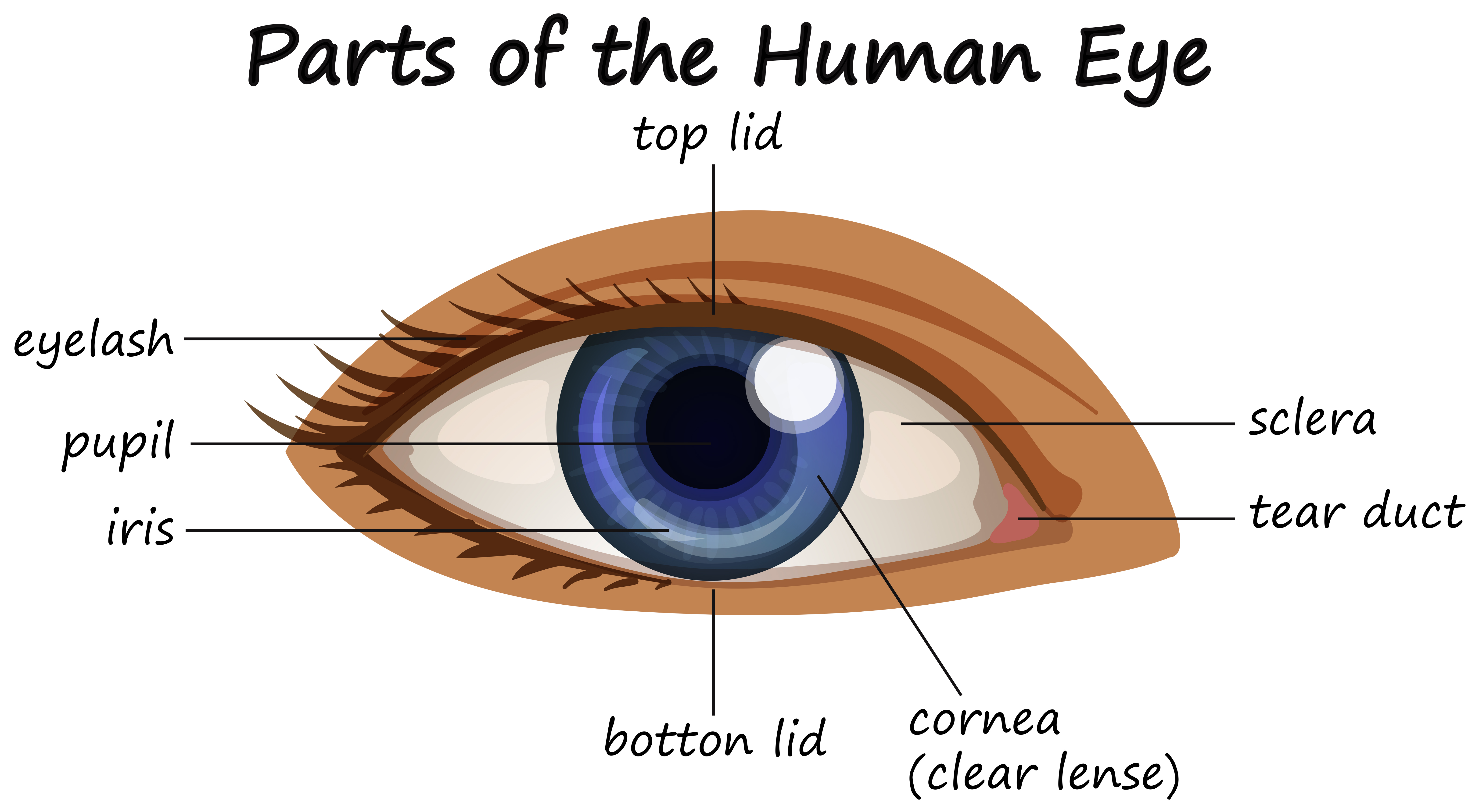
Human Eye Diagram Labeled
Download Human Eye Diagram Labeled Pics. In this image, you will find eyelid, lacrimal caruncle, tear duct, lateral rectus muscle, sclera, choroid, retina, macula lutea, fovea centralis, optic nerve and retinal blood, medial rectus muscle, ora serrata, ciliary body and muscle, suspensory ligaments, posterior chamber. Instead, it is made up of two separate segments fused together.

Instead, it is made up of two separate segments fused together.
Contrary to popular belief, the eyes are not perfectly spherical; Anterior horns of lateral ventricle. As a sense organ, the mammalian eye allows vision. Eye color, or more correctly, iris color is due to variable amounts of eumelanin (brown/black melanins) and pheomelanin (red/yellow melanins) produced by melanocytes.
Belum ada Komentar untuk "Human Eye Diagram Labeled"
Posting Komentar